Microscope Smooth Muscle Diagram - The Muscular System on emaze - Illustration about smooth muscle tissue.. Some muscles (skeletal muscles) will not contract unless stimulated by neurons; Illustration about smooth muscle tissue. Smooth muscles are usually less metabolically active and so not as dark/red as skeletal muscles. As indicated by its name, the tissue displays no striations or other distinct patterns under the microscope. Smooth muscle has a fusiform shape, which resembles a football or spindle.
The fuzz that's obvious on. Smooth muscle tissue images stock photos vectors shutterstock. Cardiac muscle action potential diagram explained. Smooth muscle cells are a lot smaller than cardiac muscle cells, and they do not branch or connect end to under the microscope, arterioles can be identified by a distinctive arrangement of endothelial cells and images for smooth muscle tissue labeled diagram world of reference. Smooth muscles are usually less metabolically active and so not as dark/red as skeletal muscles.
Muscular System - HSC PDHPE from www.pdhpe.net There are 3 different types of muscle: Instead, they have bundles of thin and have you noticed that when you look at something under a microscope it can be very confusing, but once you look at a reference diagram or picture. 3 smooth muscle microscope stock vector art and graphics. Anatomy of a relaxed and contracted smooth muscle cell. The trichome stain can be used to highlight smooth muscle cells (red) and background collagen (blue) in cases of spindled cell tumors. Smooth muscle (also known as visceral muscle due to the locations in which they are present ) is one of the three main types of muscle tissue that exist in the human body. In this scanning electron microscope image (approximately 400x) you can see the surface of a skeletal muscle fiber. Smooth muscle has a fusiform shape, which resembles a football or spindle.
Vsmcs display diversity in function and phenotype depending on their location within the arterial tree (large conduit vs.
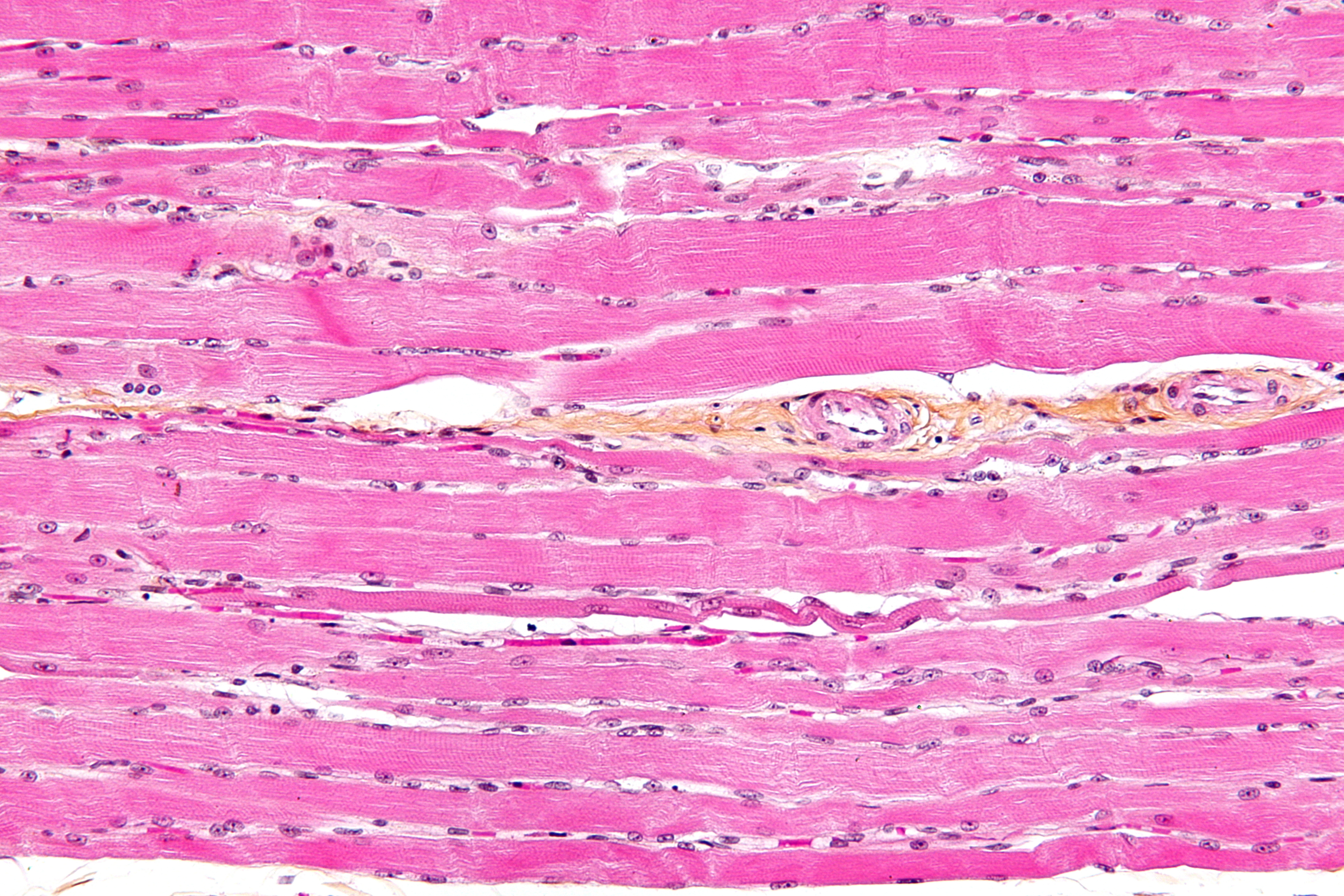
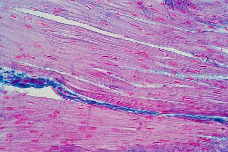
Smooth muscle (also known as visceral muscle due to the locations in which they are present ) is one of the three main types of muscle tissue that exist in the human body. Light microscopic photographs of cardiac muscle tissue with 40x. Microscopic anatomy and organization of muscle tissue. Light microscope slides of muscle tissues including cardiac muscle smoothe muscle and skeletal muscle. This page describes smooth muscle development, descriptions of cardiac muscle and smooth muscle development can be found in other notes. Anatomy of a relaxed and contracted smooth muscle cell. Fibers of smooth muscle group in branching bundles, which allows for cells to contract much stronger than those of striated musculature. Microscopic structure of muscle diagrams label the type of muscle (either skeletal, cardiac, or visceral) pictured. The smith, a mighty man is he with large and sinewy hands. How does smooth muscle tissue look under the microscope. Compared to skeletal muscle, smooth muscle cells are small. Vsmcs display diversity in function and phenotype depending on their location within the arterial tree (large conduit vs. Smooth muscle, muscle that shows no cross stripes under microscopic magnification.
Smooth muscle tissue images stock photos vectors shutterstock. This is different from cardiac muscle tissue, which develops into an as you look at this diagram of a smooth muscle fiber, you'll notice the single nucleus in the center. This is also called as histology. Download a free preview or high quality adobe illustrator ai, eps, pdf and high resolution jpeg. Smooth muscle, muscle that shows no cross stripes under microscopic magnification.
Urine: an untapped resource for human disease modeling ... from blogs.biomedcentral.com Illustration about smooth muscle tissue. Professor susan anderson helps you recognise and understand the similarities and differences between the microscopic appearances of skeletal, cardiac and smooth muscle. However, the nuclear membrane, nuclear pore Smooth muscle, muscle that shows no cross stripes under microscopic magnification. Download a free preview or high quality adobe illustrator ai, eps, pdf and high resolution jpeg. Vsmcs display diversity in function and phenotype depending on their location within the arterial tree (large conduit vs. Smooth muscle cells are a lot smaller than cardiac muscle cells, and they do not branch or connect end to under the microscope, arterioles can be identified by a distinctive arrangement of endothelial cells and images for smooth muscle tissue labeled diagram world of reference. Flat vector illustration isolated on white background.
Smooth muscle (shown at left) is found in walls of hollow organs such as the stomach.
Vsmcs display diversity in function and phenotype depending on their location within the arterial tree (large conduit vs. Smooth muscle tissue, unlike striated muscle, contracts slowly and automatically. Vascular smooth muscle cells (vsmcs) are the stromal cells of the vascular wall and are responsible for regulating arterial tone, blood pressure, and blood supply of the tissues. Microscopic structure of muscle diagrams label the type of muscle (either skeletal, cardiac, or visceral) pictured. However, the nuclear membrane, nuclear pore Anatomy of a relaxed and contracted smooth muscle cell. 3 smooth muscle microscope stock vector art and graphics. Smooth muscle is under involuntary control and is innervated by the autonomic nervous system. Microscope slides of muscle tissue; Smooth muscle (shown at left) is found in walls of hollow organs such as the stomach. The smith, a mighty man is he with large and sinewy hands. Download a free preview or high quality adobe illustrator ai, eps, pdf and high resolution jpeg. The image is available for download in high resolution quality up to 5412x3840.
In regional anatomy you study say head. This is also called as histology. Vascular smooth muscle cells (vsmcs) are the stromal cells of the vascular wall and are responsible for regulating arterial tone, blood pressure, and blood supply of the tissues. The fuzz that's obvious on. Compared to skeletal muscle, smooth muscle cells are small.
Histology Of Human Smooth Muscle Under Microscope View For ... from thumbs.dreamstime.com It constitutes much of the musculature of. However, the nuclear membrane, nuclear pore Smooth muscle is found in the wall of hollow organs, passageways, tracts, eye and skin. In this scanning electron microscope image (approximately 400x) you can see the surface of a skeletal muscle fiber. Light microscope slides of muscle tissues including cardiac muscle smoothe muscle and skeletal muscle. They also aren't as striped at the microscopic in microscopic anatomy you study the structure as seen through microscope. Flat vector illustration isolated on white background. What does your heart muscle look like down the microscope?
What does your heart muscle look like down the microscope?
However, the nuclear membrane, nuclear pore They are spindle shaped and have no striations. Smooth muscle, muscle that shows no cross stripes under microscopic magnification. The trichome stain can be used to highlight smooth muscle cells (red) and background collagen (blue) in cases of spindled cell tumors. In regional anatomy you study say head. It constitutes much of the musculature of. There are 3 different types of muscle: Find the perfect smooth muscle microscope stock illustrations from getty images. Cardiac muscle action potential diagram explained. This is also called as histology. This is different from cardiac muscle tissue, which develops into an as you look at this diagram of a smooth muscle fiber, you'll notice the single nucleus in the center. Smooth, cardiac, and skeletal muscle under microscope. Icon of smooth muscle cell under microscope.
Download a free preview or high quality adobe illustrator ai, eps, pdf and high resolution jpeg smooth muscle diagram. Smooth muscle has a fusiform shape, which resembles a football or spindle.


0 Komentar